By Phone
| Location | Phone Number |
| Illinois | 217-757-6565 |
| Wisconsin | 920-459-5171 |
Use MyHSHS
- Log in to MyHSHS.
- Select "Visits."
- Select "Request an Appointment."
- Select "Mammogram."
Regular mammograms are one of the best ways to detect breast cancer early. And when we catch breast cancer early, it’s the most treatable. Think of your mammogram as taking time for you - because you’re important.


HSHS joins the American Cancer Society in recommending women aged 40 and older of average risk have a screening mammogram every year.
You may need to begin mammograms earlier than age 40 if you have:
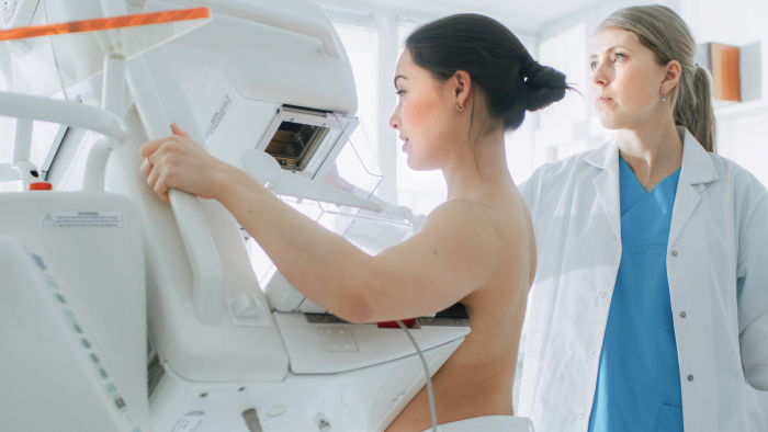
There are two main types of mammograms for breast cancer screening - 2D and 3D mammograms.
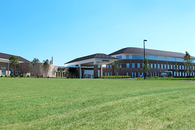
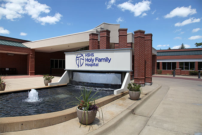
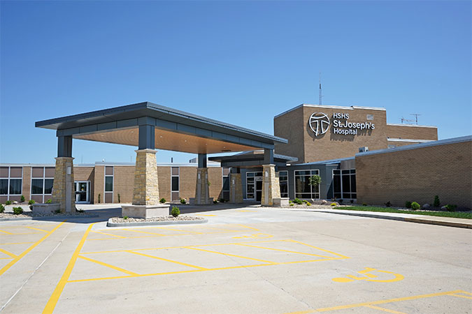
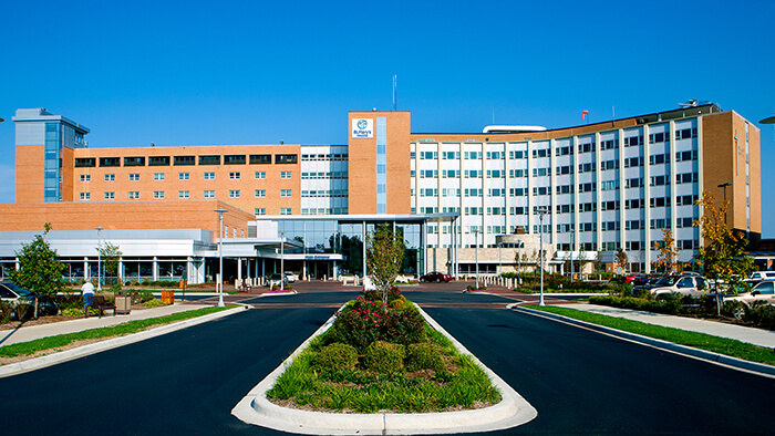
You have access to the latest in breast cancer screening and detection with 3D mammography technology near you at our HSHS locations in Illinois and Wisconsin.
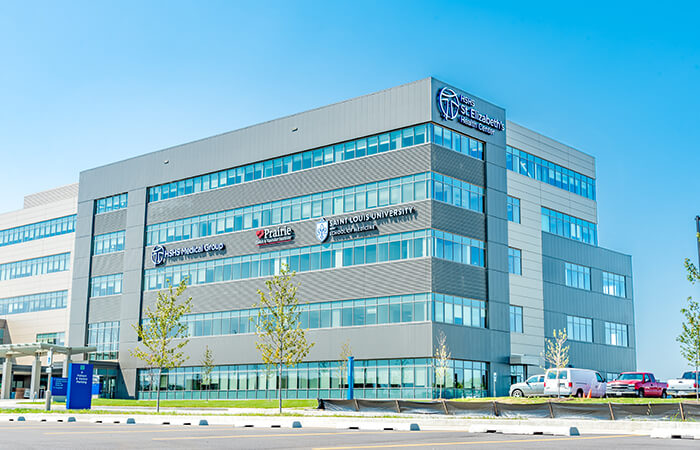
3 St. Elizabeth's Boulevard Suite 5000 O'Fallon, Illinois 62269
Mon - Thu 7:30 am - 4 pm | Fri 7:30 am - 3:30 pm
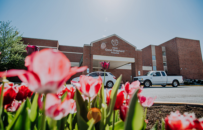
1512 N Green Mount Rd O'Fallon, Illinois 62269
400 N. 9th St. Springfield, IL 62702
1100 E Lincolnshire Boulevard Springfield, IL 62703
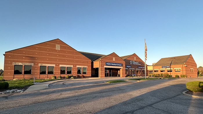
900 W Temple Ave Effingham, IL 62401
Daily | 8 am to 6 pm*
101 Coles Centre Drive, Suite 101 Mattoon, IL 61938
Monday-Friday | 8 am to 4:30 pm
1215 Franciscan Dr Litchfield, Illinois 62056-1778