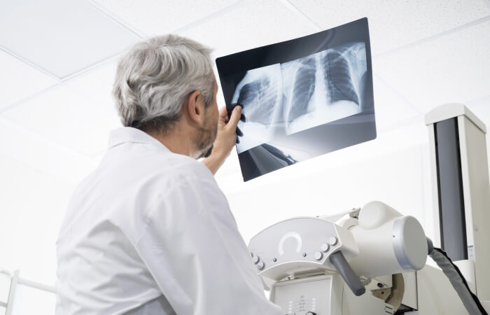
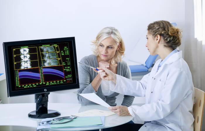
Imaging Services
Whether your physician refers you to us for an MRI, PET scan, nuclear medicine, CT scan, interventional radiology or other imaging service, rest assured that our diagnostic imaging department will provide you with high quality care.
Imaging services
HSHS St. Nicholas Hospital provides a variety of imaging services

Axumin® to detect recurrence of prostate cancer
HSHS St. Nicholas Hospital Cancer Services is now utilizing a new, state-of-the-art testing technique that helps doctors detect a recurrence of prostate cancer and save lives, better than ever before. The testing is done with an FDA-approved imaging agent called Axumin®, in combination with a positron emission tomography/Computed Tomography (PET/CT)
Prior to a PET /CT scan, the imaging agent, Axumin (also known as a “tracer”), is administered to the patient through an IV. Axumin contains a synthetic amino acid that is absorbed by prostate cancer in the body at a much more rapid pace than normal cells. Like many imaging tracers, it also contains a radioactive element which allows for Axumin to be seen inside the patient’s body during a PET/CT scan. Therefore, if there is a recurrence or metastasis of cancer in the body, the physician will be able to identify the location and extent of the cancer based on where the Axumin fluid collects in greater amounts in the body. Over time and through a natural process, the tracer will become non-radioactive, and much of it will leave your body in your urine.

Cardiac Stress Test
To prepare for your stress test, do not ingest caffeine for 24 hours prior to the test. This includes coffee, tea, soda, chocolate and medications such as Excedrin. In addition, do not eat or drink anything for four hours prior to the test. If you have questions about the test, please consult your physician.
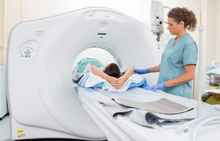
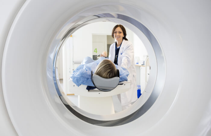
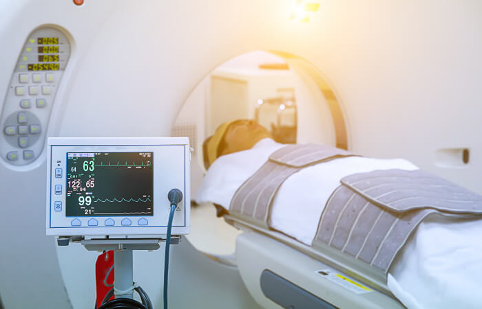
CT scan (computerized tomography)
To prepare for a CT scan
Wear loose, comfortable clothing free of metal snaps and zippers, if possible. When you arrive, you will be asked to remove metallic jewelry, watches, hair pins, hearing aids, removable dental work and glasses if they are in the area being scanned.
If you are having a CT scan of the abdominal area, you may need to drink a liquid contrast. This will be given to you, with instructions, in advance. You may be asked to not eat or drink anything for four hours before your scan. Your healthcare provider will instruct you which medications, if any, cannot be taken.
The scan typically takes 30 minutes or less. You will be asked to hold very still in specific positions as instructed by a technologist. You may be asked to hold your breath during some scans to reduce blurring on the images. If your scan requires the use of a contrast, you may be given another contrast material during the scan by IV injection. This may make you feel warm inside, but the sensation only lasts a few moments and is not painful. After the scan, you will be asked to drink plenty of fluids.
Heart Calcium Scoring
The test is performed with a cardiac CT scan to identify the presence, location and amount of calcified plaque in the coronary arteries. Calcified plaque can signal the presence of atherosclerosis, a disease of the vessel wall known as coronary artery disease. People with this disease have an increased risk for heart attack.
In general, men over the age of 30 and women over the age of 40 should consider having a heart calcium scoring. Anyone with the following risk factors should also consider heart calcium scoring:
- High cholesterol levels
- Family history of heart disease
- Diabetes
- High blood pressure
- Cigarette smoking
- Overweight or obese
- Physically inactive
Interventional Radiology
Preparation for this scan varies by patient based on the test being conducted. To learn more, visit http://www.greenbayradiology.com.
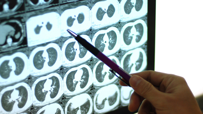
Lung cancer screening
Proven to save lives. Our screening helps catch lung cancer early before symptoms start.
Lung cancer is one of the most common forms of cancer in the United States and the leading cause of cancer death. Its symptoms often appear when the cancer is advanced and more challenging to successfully treat. However, a Low-Dose CT scan works to detect lung cancer.
What is a Low-Dose CT Lung Screening?
This is a type of X-ray that produces detailed images of your lungs with limited radiation.
Eligible participants
Low-Dose CT lung cancer screening is available for high-risk individuals with a long history of smoking. To be eligible, you must be between the ages of 55 and 77 with 30 or more pack years of cigarette smoking or meet the specific criteria set by your insurance plan.
Having 30 pack years is equivalent to smoking:
- 2 packs per day for 15 years.
- 1 pack per day for 30 years.
- ½ pack per day for 60 years.
Do I need a referral?
A referral is necessary. Please discuss this with your primary care provider to find out if this screening is right for you.
Insurance coverage
Some insurance plans do not cover this procedure. Payment through your health savings account (HAS), cash, check or credit card will be requested at the time of screening.
Mammograms
Monday - Friday 8 a.m. to 7 p.m.
We offer comprehensive breast health services within the hospital, and our mammography program is accredited by the American College of Radiology.
MRI (magnetic resonance imaging)
To prepare for an MRI:
For your safety, you will be asked to put on a patient gown for the test. Clothing with metal or pockets will not be allowed in the scanning room. Certain types of metal in the area being scanned can cause significant errors, called artifacts, in the images. It is important that metal objects are not brought into the scanning room. You will be asked to remove your jewelry, watch, hairpins, bobby pins and hearing aids. A typical exam lasts between 30 to 60 minutes for each body area being scanned. You should allow extra time because the exam may last longer than expected.
The magnet makes a “knocking” sound as images are being taken. In between scans, the machine is quiet. The MRI technologist will provide you with either ear plugs or headphones for you to listen to music during your exam.

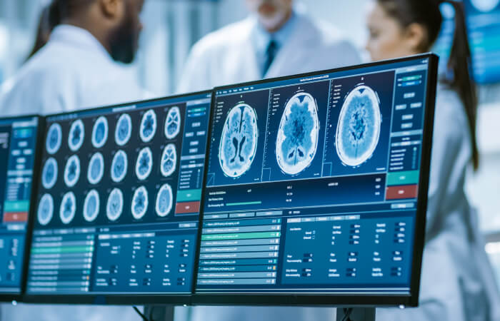
Nuclear Medicine
Depending on the type of nuclear medicine exam, the radiotracer is injected into the body, swallowed or inhaled, and accumulates in the organ or area of the body being examined. Radioactive emissions from the radiotracer are detected by a special camera that produces pictures and provides information. Scanning can begin immediately after injection, or be delayed for several hours or even days. Scan time also varies, with some scans as short as 30 minutes and others several hours. Preparation for nuclear medicine exams varies, so instructions should be reviewed and followed carefully for each exam.
PET/CT
The advantage of CT is its ability to take cross-sectional images of your body. The PET exam pinpoints metabolic activity in cells and the CT exam provides an anatomical reference. When these two scans are fused together, your physician can view metabolic changes in the proper anatomical context of your body.
To prepare for a PET/CT scan:
When you arrive for your appointment, we will review your history and any past exams. For the PET portion of the exam, you will receive a tracer material injection. For most studies, you will wait for the radiopharmaceutical to distribute itself in your body, typically 45 minutes to an hour. You are asked to relax and no reading or TV is permitted. Then, during the exam, you will lie very still on a comfortable table that will move slowly through the scanner as it acquires the information needed to generate diagnostic images. The PET/CT scan itself should last about 45 minutes, but the exam procedure can vary depending on what we are looking for and what we discover along the way. Plan to spend two to three hours with us.
Our PET/CT scans are accredited by the Intersocietal Accreditation Commission.
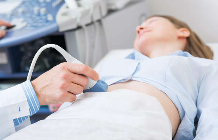
Ultrasound
Ultrasound at HSHS St. Nicholas Hospital is accredited by the American College of Radiology (ACR).