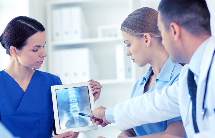
Neuroscience Technology
The HSHS St. Vincent Hospital Neuroscience Center uses leading-edge technology to guide their surgical planning and ensure the highest level of precision when patients need it most.
Spine Robot
The spine robot is designed to improve accuracy and optimize patient care by using robotics and navigation, much like a GPS in your car. On the day of surgery, medical images are taken and imported into the machine. The surgeon uses these images to determine the size and placement of implants and creates a patient plan based on the anatomy. This is used to guide the rigid robotic arm to a specific region of your spine, similar to a planned route or pathway on a GPS. The surgeon uses this pathway or route to accurately place the implants using instruments. Throughout the procedure, the surgical instruments and implants are continuously displayed on the screen for the surgeon and staff to monitor. This display allows the surgeon to view live feedback during the procedure for more precise implant placement.
Biplane Angiography Technology and Thrombectomy
Biplane angiography is a medical imaging technology used primarily in interventional radiology and cardiology procedures. This advanced imaging technique provides real-time, high-quality images of blood vessels, helping medical professionals visualize and diagnose various vascular conditions. The term "biplane" refers to the use of two separate X-ray imaging systems, positioned at right angles to each other, to capture images from different perspectives simultaneously.
Thrombectomy is a neurointerventional procedure that removes a blood clot, or thrombus, from a blood vessel. This procedure is commonly used in the life-saving treatment of a stroke, where a blood clot obstructs the brain's blood flow. By removing the blood clot, normal blood flow is restored to prevent further damage to the brain.
The combination of thrombectomy and biplane imaging allows medical professionals to see the blood vessels in multiple dimensions, improving the ability to navigate and precisely place devices that perform the thrombectomy to quickly extract blood clots – all while continuously monitoring the patient. The real-time visualization provided by biplane imaging assists healthcare professionals in performing the thrombectomy accurately, efficiently and promptly.
Gleolan
High-grade gliomas, such as glioblastomas, infiltrate into a person’s brain tissue which can make it difficult for surgeons to differentiate between cancerous and non-cancerous tissue in the brain during surgery. In some cases, the cancer appears to disguise itself as healthy brain tissue.
Gleolan is an imaging agent that assists the surgeon in pinpointing where the cancerous tissue is located within the brain. Patients ingest the Gleolan solution 2 to 4 hours prior to the surgery. Then, during surgery, the surgeon views the brain through a special blue light filter on a surgical microscope. Under this blue light, the active substance in Gleolan causes the cancerous tumor cells to appear a red-violet color, while the non-cancerous brain cells appear blue. This differentiation provides the surgeon a better view, allowing them to remove more of the tumor and less healthy brain tissue. The goal is to remove as much of the tumor as possible without harming areas of the brain that control critical functions such as speech, balance, movement, memory, understanding and vision.
Stealth computer navigation
Stealth Computer Navigation is a navigation system that provides the surgeon information, via CT and MRI scans taken prior to surgery, to guide surgical planning and the approach they will use for surgery. These scans are uploaded to a computer and allow the surgeon to pinpoint the exact location of the issue. It is like a GPS system of sorts, in that the surgeon can see a 3D image of the patient’s anatomy before and during the procedure, while also seeing the exact location of their surgical instruments via two cameras.
The Stealth system is used in a variety of cranial procedures, including tumor resections, brain biopsies and shunt placements. There are many advantages to using the Stealth system — more precise localization of tumors, more complete resection, shorter surgery times and shorter hospital stays.
O-arm Surgical Imaging System
The O-arm is a surgical imaging system used in spinal and spinal trauma-related surgeries. It provides real-time intraoperative 2D and 3D images of the anatomy. The O-arm is used in conjunction with the Stealth system to provide the surgeon with improved visualization when placing spinal instrumentation. It also allows for the confirmation of surgical accuracy before the patient leaves the operating room. This technology helps to reduce complications and improve patient outcomes during spine surgery.
Stereotactic Radiosurgery (SRS)
Stereotactic Radiosurgery (SRS) is not surgery, but rather a form of radiation therapy used to treat some brain tumors and brain abnormalities (i.e. AVMs- a tangle of expanded blood vessels which can impede normal blood flow in the brain). SRS focuses high-power energy on small areas in the brain. In doing so, the treatment can be very precise and less surrounding healthy tissue gets damaged. Patients will need fewer doses than traditional radiation therapy. SRS may be a good option for patients who are too high risk for surgery whether it be related to age, medical problems, or the location of the brain tumor or abnormality. SRS doesn't remove the actual brain tumor or abnormality; instead, it damages the DNA of the tumor cells so they can no longer reproduce. It causes the blood vessels in AVMs to thicken and eventually close off so they no longer disrupt the normal blood flow. SRS can usually be done on an outpatient basis.