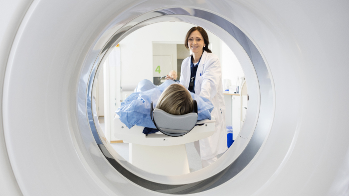
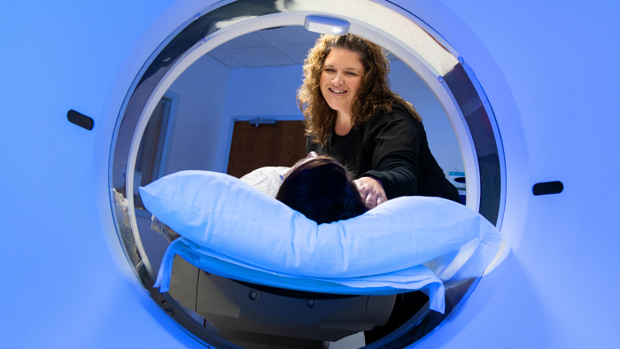
Computed Tomography (CT or CAT scan) is a non-invasive procedure that enables medical professionals to obtain accurate diagnostic information on many areas of your body. The CT uses x-rays to produce a cross-sectional image (or slice) of the areas of your body being examined. Our state-of-the-art CT scanners are capable of capturing entire organs in seconds. They provide outstanding image detail for any exam, including cardiac, head, spine, and abdominal and vascular studies.
Physicians use CT scans to examine injuries as well as diagnose and treat diseases and conditions. This can include:
- Helping to guide biopsies and other cancer treatments
- Planning and following up on surgery
- Staging and planning radiation
- Monitoring chemotherapy
- Examining injuries to internal organs and vessels
This technology allows us to image smaller structures with great detail and resolution in even the most challenging applications. CT captured images give your doctor an unprecedented level of detail needed to treat disease and life-threatening illnesses. CT at St. Joseph's Hospital gives:
- Greater diagnostic confidence
- Faster scans
- Optimized radiation doses
- Shorter breath holds for less discomfort
- Non-invasive exams with no recovery time
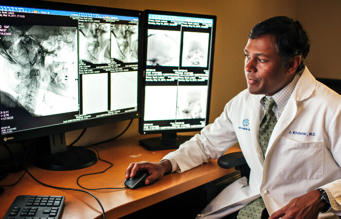
The radiologist will review the images from your CT scan and provide a detailed report to your physician. Your physician will discuss these results with you and explain what they mean, relative to your health.