Imaging Services
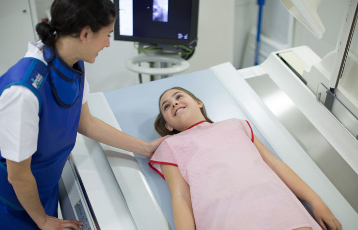
Our radiology department has the tools and techniques needed to find out what’s going on inside your child’s body when they’re sick or hurt. We care for infants, children and adolescents through the diagnosis of disease using various imaging techniques.
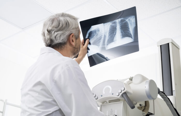
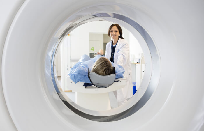
CT scan (computerized tomography)
The scanner is used in combination with a digital computer to create "slices" of different organs of the body, making it possible to detect diseases sooner than with a regular X-ray.
To prepare for a CT scan
We understand that medical imaging can feel new and unfamiliar for children. That's why we offer the “Kitten Scanner,” a child-sized practice scanner that helps kids prepare for their CT scan. Children can send a stuffed animal companion (Ollie the Elephant, Chris the Alligator or Doris the Chicken) through the toy scanner, which mimics the lights, sounds and process of a real CT scan. This playful introduction helps reduce anxiety by making the procedure familiar with a cozy friend. While sedation is available, practice through play often allows children to remain calm and still during their scan without medication.
Children should wear loose, comfortable clothing free of metal snaps and zippers, if possible. When you arrive, your child will be asked to remove metallic jewelry, watches, hair pins, hearing aids, removable dental work and glasses if they are in the area being scanned.
What to expect
- A typical scan takes 30 minutes or less.
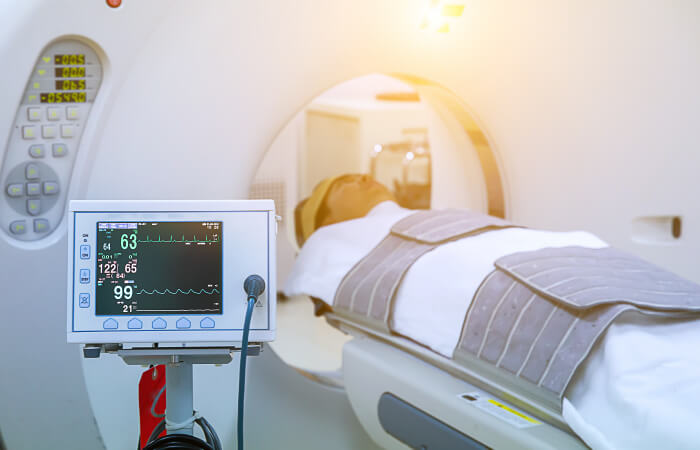
- During this test, the patient lies in a doughnut-shaped machine that takes pictures of the body.
- If your child is having a CT scan of the abdominal area, they may need to drink a liquid contrast. This will be given to them, with instructions, in advance. Your child may be asked to not eat or drink anything for four hours before their scan. Your child's healthcare provider will instruct you which medications, if any, cannot be taken.
- Your child will be asked to hold very still in specific positions as instructed by a technologist. Your child may be asked to hold their breath during some scans to reduce blurring on the images. If the scan requires the use of a contrast, your child may be given another contrast material during the scan by IV injection. This may make your child feel warm inside, but the sensation only lasts a few moments and is not painful.
- After the scan, your child will be asked to drink plenty of fluids.
The CT scanner at HSHS St. Vincent Children’s Hospital is American College of Radiology (ACR) accredited.
Interventional Radiology
Preparation for this scan varies by patient based on the test being conducted. To learn more, visit http://www.greenbayradiology.com.
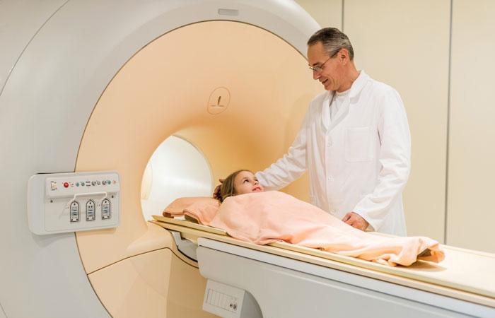
MRI (magnetic resonance imaging)
An MRI is a non-invasive and painless procedure in which radio waves and powerful magnets linked to a computer are used to create remarkably clear and detailed pictures of internal organs and tissues without the use of radiation.
To prepare for an MRI
We understand that medical imaging can feel new and unfamiliar for children. That's why we offer the “Kitten Scanner,” a child-sized practice scanner that helps kids prepare for their MRI. Children can send a stuffed animal companion (Ollie the Elephant, Chris the Alligator or Doris the Chicken) through the toy scanner, which mimics the lights, sounds and process of a real MRI. This playful introduction helps reduce anxiety by making the procedure familiar with a cozy friend. While sedation is available, practice through play often allows children to remain calm and still during their scan without medication.
What to expect
- A typical exam lasts between 30 to 60 minutes for each body area being scanned. You should allow extra time because the exam may last longer than expected.
- For your child's safety, they will be asked to put on a patient gown for the test as clothing with metal or pockets will not be allowed in the scanning room. Certain types of metal in the area being scanned can cause significant errors, called artifacts, in the images. Your child will also be asked to remove their jewelry, watch, hairpins, bobby pins and hearing aids. and It is important that metal objects are not brought into the scanning room.
- The magnet makes a "knocking" sound as images are being taken. In between scans, the machine is quiet. The MRI technologist will provide your child with ear plugs or headphones to listen to music during their exam.
MRI at HSHS St. Vincent Children’s Hospital is American College of Radiology (ACR) accredited.
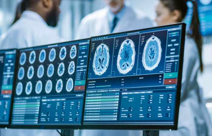
PET/CT (Positron emission tomography)
This procedure combines functional information from a Positron Emission Tomography (PET) exam with anatomical information from a computed tomography (CT) scan.
The advantage of CT is its ability to take cross-sectional images of your child's body. The PET exam pinpoints metabolic activity in cells and the CT exam provides an anatomical reference. When these two scans are fused together, your child's physician can view metabolic changes in the proper anatomical context of their body.
To prepare for a PET/CT scan
We understand that medical imaging can feel new and unfamiliar for children. That's why we offer the “Kitten Scanner,” a child-sized practice scanner that helps kids prepare for the CT portion of their PET/CT procedure. Children can send a stuffed animal companion (Ollie the Elephant, Chris the Alligator or Doris the Chicken) through the toy scanner, which mimics the lights, sounds and process of a real CT scan. This playful introduction helps reduce anxiety by making the procedure familiar with a cozy friend. While sedation is available, practice through play often allows children to remain calm and still during their scan without medication.
What to expect
- The PET/CT scan itself should last about 45 minutes, but the exam procedure can vary depending on what we are looking for and what we discover along the way. Plan to spend two to three hours with us.
- When you arrive for your child's appointment, we will review your child's history and any past exams.
- For the PET portion of the procedure, your child will receive a tracer material injection. For most studies, your child will wait for the radiopharmaceutical to distribute itself in their body, typically 45 minutes to an hour. Your child will be asked to relax and no reading or TV is permitted.
- Then, during the CT scan, your child will lie very still on a comfortable table that will move slowly through the scanner as it acquires the information needed to generate diagnostic images.
Our PET/CT scans are accredited by the Intersocietal Accreditation Commission.
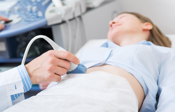
Ultrasound
Ultrasound at HSHS St. Vincent Hospital is accredited by ACR.