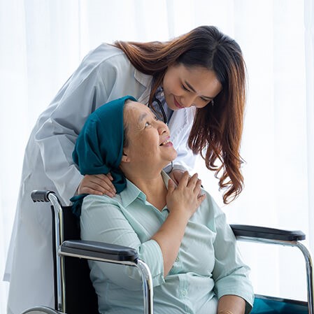
At HSHS St. Mary’s Hospital Cancer Care Center, our team welcomes you with open arms. Jenny, a young mother recently diagnosed with cancer, shares her message of gratitude with her caregivers in a letter she wrote to the cancer care team.
Cancer Care
The Cancer Care Center at HSHS St. Mary’s Hospital combines state-of-the-art oncology treatment technology with compassionate care for your body and soul.

Treatment Options
We provide radiation as well as chemotherapy treatment, and we can treat almost any type of cancer regardless of diagnosis or stage. In addition, we provide TomoTherapy Hi-Art radiation treatment which offers an unmatched level of accuracy and minimizes side effects typically associated with previous radiation therapy methods.
A Team of Experts
Meet your exceptional cancer care team, led by specialists from The Cancer Care Specialists of Central Illinois. Radiation oncologists, medical oncologists and hematologists work together with the Cancer Care Center's team of medical physicists, dosimetrists, nurses, radiation therapists and administrative colleagues to develop and monitor a treatment plan to meet your needs.
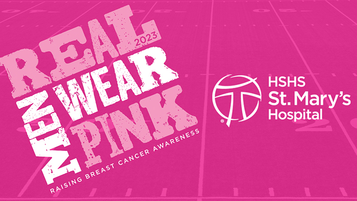
Real Men Wear Pink
The goal of the RMWP campaign is to generate breast cancer awareness and to encourage our mothers, aunts, grandmothers, sisters and loved ones to meet with their doctor to discuss breast cancer screenings.
Funds raised in the Real Men Wear Pink Campaign go toward breast cancer awareness and the HSHS St. Mary' Hospital Cancer Care Center. Click here to make a donation to the HSHS St. Mary's Foundation.