Diagnostic Imaging

HSHS St. Mary's Hospital Radiology Services offers the latest in health care technology to patients and physicians in our region.

Imaging doctor reviewing imagery from lab results and testing

With this technology, those who come to our hospital are assured they receive the most advanced care close to home.

Our talented physicians and team use this technology to accurately identify fractures, ultrasounds for expectant mothers, and screening life-threatening diseases, such as cancer, breast cancer, heart disease, and Alzheimer's disease at earlier stages, providing hope and saving lives.

To schedule diagnostic imaging services, please call 217-464-2984.

Patient in CT scanner with technician assisting

CT Scans

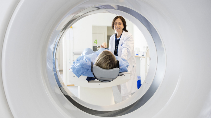
Computed Tomography (CT or CAT scan) is a non-invasive procedure that enables medical professionals to obtain accurate diagnostic information on many areas of your body. The CT uses x-rays to produce a cross-sectional image (or slice) of the areas of your body being examined. Our state-of-the-art CT scanners are capable of capturing entire organs in seconds. They provide outstanding image detail for any exam, including cardiac, head, spine, and abdominal and vascular studies.

This technology allows us to image smaller structures with great detail and resolution in even the most challenging applications. CT captured images give your doctor an unprecedented level of detail needed to treat disease and life-threatening illnesses. CT at St. Elizabeth’s Hospital gives:

  • Greater diagnostic confidence
  • Faster scans
  • Optimized radiation doses
  • Shorter breath holds for less discomfort
  • Non-invasive exams with no recovery time

As a patient:

  • If instructed, drink contrast (a liquid that improves the image) 1 1/2 hours before your CT.
  • The test may take 10 minutes to 2 hours, depending on whether contrast is used and the part of the body being scanned.
  • Arrive on time to check in, you may be asked to change into a hospital gown.
  • You may be given contrast through an intravenous (IV) line or by injection.
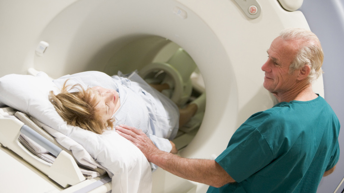
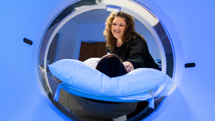
  • You will lie on a table. The table slides into the CT scanner.
  • The technologist will ask you to hold your breath for a few seconds during your scan.
  • After your test, you can go back to your normal diet and activities right away. Any contrast will pass naturally through your body within a day.


Patient in an MRI machine with technician looking on

MRI

St. Elizabeth’s Hospital offers the cutting-edge 1.5T magnetic resonance imaging (MRI) system. The system is the quietest MRI on the market to the extent that there is no ear protection necessary for maximum patient comfort. The 1.5T MRI delivers high-speed, high-resolution imaging that produces accurate and reliable results with unmatched image quality. The new system design features superior coil technology that allows clinicians to perform multiple exams without repositioning the patient.

In comparison to other higher magnetic resonance machines, the patient friendly 1.5T offers significant advantages including wide frame opening and larger cabin space. The magnet is housed in a comfortable, relaxing environment designed to minimize patient anxiety.The MRI capabilities include non-contrast MRA technique, a method of non-invasive angiography that images the arterial and/or venous system without contrast injection. This is especially useful for patients with decreased renal function because of direct side effects caused by contrast injections. The system also offers sophisticated breast imaging and guidance for breast biopsy.

As a patient:

  • MRI uses strong magnets, so you will be asked to remove your watch, jewelry, and other metal objects.
  • You may be asked to remove your makeup, which may contain some metal.
  • The test takes 30-60 minutes.
  • You may be injected with contrast (a special “dye” that improves the MRI image).
  • You will lie down on a platform that slides into the magnet.
  • After your test, you can get back to normal activities right away. If you were given contrast, it will pass naturally through your body within a day.

Nuclear Medicine

Nuclear medicine is a radiology subspecialty using trace amounts of radioactive material to diagnose and treat many diseases. Nuclear imaging does not require surgery. Instead, it relies on radioactive drugs or radiotracers.

The radiotracer can be injected, swallowed or inhaled as a gas, depending on your test. It gives off gamma rays, which are detected by a Nuclear Medicine scanner, a special camera or a probe. Using a computer, your health care team will measure the amount of the radiotracer absorbed by the body to produce images offering details of your body to help in your diagnosis and treatment.

At St. Mary's Hospital, Nuclear Medicine is used to:

  • Analyze the functions of organisms such as kidney, gallbladder and lungs
  • Check bones for fractures, tumors, etc.
  • Visualize blood flow
  • Monitor presence or spread of cancer
  • Locate infection in the body

As a patient:

  • You will be given a tracer (radioactive material). It may be injected, swallowed, or inhaled.
  • Your scan may then be done right away, or you may need to wait a few hours or even days to allow the tracer to concentrate in the part of the body being studied.
  • Your scan may take a few hours. Bring something you can do if you need to wait.
  • You will lie on a narrow imaging table; a large camera is placed close to your body.
  • Remain as still as you can while the camera takes the pictures to ensure the best imaging.
  • After your scan, drink plenty of water to help clear the tracer from your body.

More Imaging services

Interventional Radiology
Interventional radiology, commonly referred to as IR, is the combination of very small instruments and digital imaging. Interventional radiologic procedures use diagnostic-type imaging equipment to assist a physician in the treatment of a patient's condition. These procedures frequently provide favorable medical results with minimal recovery time. In some cases these procedures avoid the need for conventional surgery or improve the prospects for a favorable outcome from surgery. Our interventional equipment is located on 6th floor or within the Radiology Department depending on the exam scheduled.
 
Procedures Performed
  • CT and Ultrasound guided biopsies
  • Hip Injections
  • Abscess drainage procedures
  • Lumbar puncture
  • Myelography
X-Rays
X-ray, commonly referred to as radiography, uses ionizing radiation to provide images of the body. Radiography is used in many ways to diagnose disease and injuries. Some common exams are X-rays, GI fluoroscopy exams and joint injections (arthrograms). Preparation for this scan varies by patient based on the test being conducted.

 
Bone Density
Bone densitometry is most often used to diagnose osteoporosis, a condition that often affects women after menopause but may also be found in men. Osteoporosis involves a gradual loss of calcium, causing the bones to become thinner, more fragile and more likely to break.

Bone Densitometry is also effective in tracking the effects of treatment for osteoporosis and other conditions that cause bone loss. A Bone Densitometry test can also assess an individual’s risk for developing fractures.

Ask your primary care physician if you should consider having a Bone Densitometry test.

To schedule an appointment, call 217-464-2976.
Outpatient Biopsy
A biopsy is the removal of tissue in order to examine it for disease. The tissue samples can be taken from any part of the body.

Biopsies are performed in several different ways. Some biopsies involve removing a small amount of tissue with a needle while others involve surgically removing an entire lump, or nodule, that is suspicious.

Often, the tissue is removed by placing a needle through the skin to the area of abnormality. Biopsies can be safely performed with imaging guidance such as ultrasound, x-ray, or computed tomography (CT). These types of imaging are used to determine exactly where to place the needle and perform the biopsy.
Breast Ultrasound
Ultrasound, sometimes called ultrasound scanning or sonography, is safe and painless. It produces pictures of the inside of the body using sound waves.

It uses a small probe called a transducer and gel placed directly on the skin. High-frequency sound waves travel from the probe through the gel into the body. The probe collects the sounds that bounce back. A computer uses those sound waves to create an image.

Ultrasound exams do not use radiation (as used in x-rays). Because images are captured in real-time, they can show the structure and movement of the body's internal organs. They can also show blood flowing through blood vessels.

Ultrasound imaging is a noninvasive medical test that helps physicians diagnose and treat medical conditions.
Ultrasound
Ultrasound is one of the most widely used diagnostic procedures available. It provides a safe, non-invasive and virtually painless means of observing soft tissue anatomy on an outpatient basis.

Medical sonography (ultrasonography) is an ultrasound-based diagnostic medical imaging technique used to visualize muscles, tendons, and many internal organs, their size, structure and any pathological lesions with real time tomographic images. It is also used to visualize a fetus during routine and emergency prenatal care. Ultrasound scans are performed by medical healthcare professionals called sonographers. Obstetric sonography is commonly used during pregnancy.

What are other applications for Ultrasound
  • OB/GYN: evaluation of fetal anatomy, age, growth and position; imaging of the uterus and ovaries to evaluate pelvic abnormalities.
  • Radiology: identification of pathology in abdominal organs, OB/GYN and prostate scanning, evaluation of blood flow throughout the vascular system.
  • Vascular: examination of vessels, such as the carotid arteries, to detect and precisely locate any possible blood flow obstructions.
  • Musculoskeletal: examination of tendons, ligaments and joint spaces to evaluate shoulder, Achilles tendon and repetitive motion injuries. With ultrasound, the clinician can examine your shoulder or ankle, for instance, while you perform a range-of-motion movement.
  • Pediatrics: aid in detection of congenital conditions and the cardiovascular system.
As a Patient:
  • After you are positioned on the examination table, the radiologist or sonographer will apply some warm gel on your skin and then place the transducer firmly against your body, moving it back and forth over the area of interest until the desired images are captured.
  • Ultrasound exams in which the transducer is inserted into an opening of the body may produce minimal discomfort.
  • Once the imaging is complete, the gel will be wiped off your skin.
  • After an ultrasound exam, you should be able to resume your normal activities.
Are there risks or side effects?
No. The sound waves will not cause discomfort and they do not contain ionizing radiation.