Vascular Services

Non-invasive imaging technology to study arteries, veins and vessels

Man performing doppler study on woman's leg

Vascular Lab Studies uses doppler, ultrasound, and sound waves to diagnose vascular disease in a non-invasive manner. Our lab specializes in several tests that look for vascular diseases that may affect how well blood flows in the arteries and veins. The lab examines blood vessels throughout the body that feed major organs and tissue.

HSHS St. Mary’s Hospital's vascular lab enables patients to undergo basic angiography procedures—X-ray examinations of the peripheral blood vessels (vessels outside the heart)—and necessary interventional procedures in the same room. Patients benefit from rapid scan times, optimal image quality and maximum protection from radiation, as physicians view even the smallest vascular structures in detail.

Peripheral Vascular Disease (PVD)

PVD is a build-up of plaque in the arteries outside your heart that reduces the flow of blood. As a result, some parts of your body do not get the oxygen they need. People with PVD are three to four times more likely to die from stroke or heart attack than people of the same age who do not have PVD.

Symptoms include:

  • Dull, cramping pain in hips, thighs or calf muscle
  • Pain in the legs often occurs when walking or exercising and is relieved by rest
  • Buttock pain
  • Numbness or tingling in the leg, foot, or toes
  • Changes in skin color (pale, bluish, or reddish discoloration)
  • Changes in skin temperature, coolness
  • Impotence
  • Infection/sores that do not heal
  • Thickened nails

Tests to diagnose this condition include:

  • Ankle Brachial Index (ABI)  – This is a simple non-invasive test that measures the ratio of the blood pressure in your ankle to that in your arm by placing blood pressure cuffs on the arms and lower legs. This ratio may indicate a potential vascular problem.
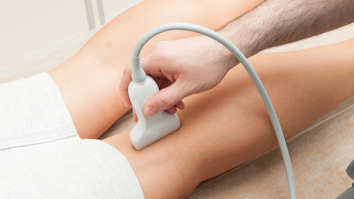
  • Ultrasound Duplex Test  – This is a non invasive test using sound waves to provide image of the inside of the blood vessel to determine if a specific artery has plaque build up. There is no radiation used and generally no discomfort from the application of the ultrasound transducer to the skin.
Woman rubbing her leg

Deep Vein Thrombosis (DVT)

DVT is a blood clot that develops in a vein deep in the body. The clot may partially or completely block blood flow through the vein. Most DVTs occur in the lower leg, thigh or pelvis, although they also can occur in other part of the body including the arm, brain, intestines, liver, or kidney.

DVT most commonly occurs in just one leg or arm. Not everyone with DVT will experience symptoms, although when present, they may include:

  • Swelling of the leg or arm (sometimes suddenly).
  • Pain or tenderness in the leg that may only be present when standing or walking.
  • Feeling of increased warmth in the area of the leg or arm that is swollen or that hurts.
  • Redness or discoloration of the skin.
  • Enlargement of the superficial veins in the affected leg or arm.

Tests to diagnose this condition include:

Duplex Venous Ultrasound  – This test is used to evaluate the blood flow in the veins and to detect the presence and specific location of blood clots. A technologist applies pressure when scanning your arm or leg. If the vein does not compress with pressure, it may indicate a blood clot is present.

Woman receiving a ultrasound on her neck

Carotid Artery Disease

Carotid Artery Disease is the build-up of plaque on the inside of the carotid arteries. The carotid arteries carry oxygen-rich blood away from the heart to the head and body. Over time, the build-up narrows the artery, decreasing blood flow to the brain which can lead to a stroke.

There may not be any symptoms of carotid artery disease. A transient ischemic attack (also called TIA or “mini-stroke”) is one of the most important warning signs of a stroke. A TIA is a temporary episode of:

  • Blurred or loss of vision in one or both eyes
  • Weakness and/or numbness of your arm, leg or face on one side of your body
  • Slurring of speech, difficulty talking or understanding what others are saying
  • Loss of coordination, dizziness or confusion
  • Trouble swallowing

Tests to diagnose this condition include:

  • Carotid Duplex Ultrasound – A procedure that uses high-frequency sound waves to view the blood vessels in the neck to determine the presence of narrowing in the carotid arteries.
  • Computerized Tomography (CT Scan)  – A CT of the brain may be performed if there is a possibility a stroke has already occurred. This test will reveal areas of damage on the brain.

Abdominal Aortic Aneurysm (AAA)

An Abdominal Aortic Aneurysm is a dilation of a segment of the aorta, the main artery through the trunk of the body.

An AAA can be detected by using ultrasound imaging which provides an accurate, safe, and non-invasive means to measure the aortic size. The most important risk factors for AAA include:

  • Age over 60 years
  • Family history of AAA
  • Smoking
  • Hypertension

Renal Artery Disease

The renal arteries provide blood flow to the kidneys and can become diseased which results in reduced blood flow to the kidneys. This reduced blood flow results in hypertension. Renal Artery Stenosis is the most common correctable cause of hypertension. Long-standing, untreated Renal Artery Disease is also an important cause of kidney failure.

Renal Artery Disease can be detected by using ultrasound imaging which is an accurate, non-invasive, and cost-effective method of diagnosing disease without using contrast material.


Preparing for your vascular test

Carotid, Arterial, and Venous Ultrasound Test

  • Take your prescribed morning medications; including your heart and/or blood pressure medications.
  • Wear comfortable clothing. If having a Carotid ultrasound, make sure to wear a shirt that is loose fitting around the neck.
  • Leave all jewelry, valuables and money at home.

Abdominal Ultrasound (Renal, Aorta and Iliac) Test

What to do the day before your vascular ultrasound test:

  • Take an antigas medication, such as Gas-X, before going to bed.
  • No food after midnight.

The day of your procedure:

  • No food the morning of the testing.
  • Take your prescribed morning medications with small sips of water.
  • If you take insulin or medicine for diabetes, wait and take after testing.