Wiesemann Women’s Center

Exceptional care and advanced technology with comfort of caring individuals for women

Woman wearing glasses smiling

HSHS St. Mary's Wiesemann Women's Center provides care to women from prevention, screening, and diagnosis to treatment and follow-up care in a consistent, compassionate and timely manner. For more information or to schedule an appointment, call 217-464-2976.

Woman laying on diagnostic table for bone density test

Bone densitometry

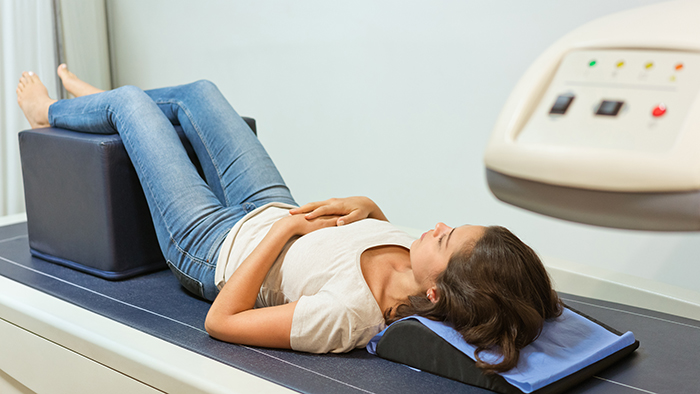
Bone densitometry is most often used to diagnose osteoporosis, a condition that often affects women after menopause but may also be found in men. Osteoporosis involves a gradual loss of calcium, causing the bones to become thinner, more fragile and more likely to break.

Bone Densitometry is also effective in tracking the effects of treatment for osteoporosis and other conditions that cause bone loss. A Bone Densitometry test can also assess an individual’s risk for developing fractures.

Ask your primary care physician if you should consider having a Bone Densitometry test.

Young woman receiving a mammography exam

Mammography

A mammogram is an x-ray examination of the breast done in complete privacy by a specially-trained female technologist.

Breast biopsies

This procedure is performed to collect a sample of cells or tissue from the suspicious area for examination under a microscope by a pathologist. We offer several alternatives to surgery, which are faster and less painful, with fewer complications and less recovery time.  Please ask our Women’s Wellness Center staff for more information.

Stereotactic Breast Biopsy

Stereotactic Breast Biopsy offers women a faster and less painful alternative to surgery, with fewer complications and less recovery time. This procedure is performed to collect a sample of cells or tissue from the suspicious area for examination under a microscope by a pathologist.

Young female doctor with pink awareness ribbon holding hands of senior woman

Breast ultrasound

Ultrasound, sometimes called ultrasound scanning or sonography, is safe and painless. It produces pictures of the inside of the body using sound waves.

It uses a small probe called a transducer and gel placed directly on the skin. High-frequency sound waves travel from the probe through the gel into the body. The probe collects the sounds that bounce back. A computer uses those sound waves to create an image.

Ultrasound exams do not use radiation (as used in x-rays). Because images are captured in real-time, they can show the structure and movement of the body's internal organs. They can also show blood flowing through blood vessels.

Ultrasound imaging is a noninvasive medical test that helps physicians diagnose and treat medical conditions.

Young woman receiving an ultrasound exam

Ultrasound

Ultrasound, also called ultrasound scanning or sonography, involves exposing part of the body to high-frequency sound waves to produce pictures of the inside of the body. Ultrasound exams do not use ionizing radiation (as used in x-rays). Because ultrasound images are captured in real-time, they can show the structure and movement of the body's internal organs, as well as blood flowing through blood vessels.

All Sonographers at St. Mary’s are registered by the ARDMS. The department has ACR accreditation for general OB/GYN studies. The department has ICAVL accreditation for all vascular studies as well. Questions? Call us at 217-464-2163.

What to expect:

  • After you are positioned on the examination table, the radiologist or sonographer will apply some warm gel on your skin and then place the transducer firmly against your body, moving it back and forth over the area of interest until the desired images are captured.
  • Ultrasound exams in which the transducer is inserted into an opening of the body may produce minimal discomfort.
  • Once the imaging is complete, the gel will be wiped off your skin.
  • After an ultrasound exam, you should be able to resume your normal activities.

Exams Performed:

  • Abdominal scanning
  • OB/GYN scanning (includes transvaginal and Biophysical Profiles)
  • Core Breast Biopsies
  • Small Parts
  • Vascular (includes carotids, venous and arterial)
  • US guided biopsies; Amniocentesis; Aspirations; Paracentesis; Thoracentesis
  • Within the hospital, Echocardiographies are also performed in the Cardiovascular Lab.