Imaging services
HSHS St. John's Imaging (Radiology) Department is staffed by expertly trained medical professionals who use state-of-the-art imaging equipment.
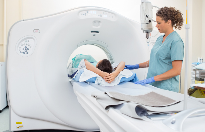
CT scan
Computed Tomography (CT or CAT) scans are non-invasive diagnostic tests that generate three-dimensional images of your internal body. CT scans create images by combining multiple “slices” of digital information into a single image that is primarily used to diagnose and treat cancers and other internal medical issues. CT scans typically focus on internal organs, bone, soft tissue, and blood vessels and have the ability to show more detail than traditional x-rays. Frequently, patients are given a contrast agent by injection or to drink prior to the exam to help to reveal details of specific areas of the body for clearer imaging.

MRI and Open MRI
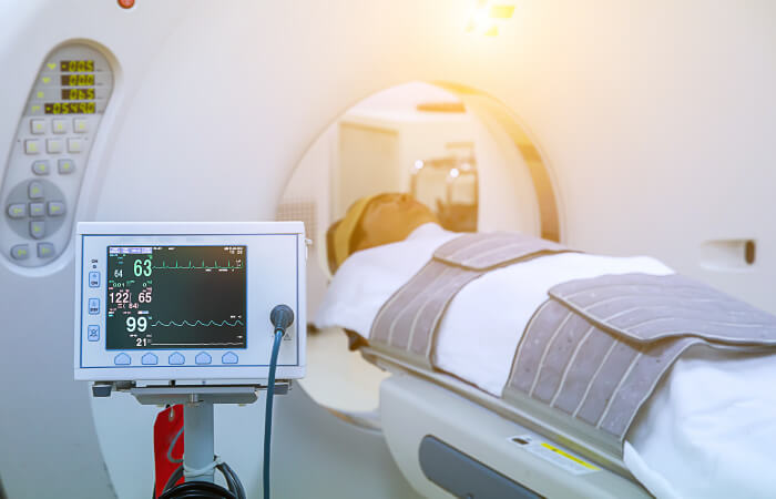
Magnetic Resonance Imaging (MRI) creates highly detailed images of your internal body. This non-invasive, diagnostic testing procedure offers the most detailed images of soft tissues and organs. It is especially effective in the evaluation of neuromuscular disorders such as brain abnormalities, back pain, and joint injuries. MRI excels in detection of breast cancer, as well as body solid organ diseases such as liver and kidney problems. In addition, MRI procedures do not employ radiation that is used in traditional x-rays and CT scans.
A high strength, or high field, MRI offers the highest resolution images possible with unmatched speed. These detailed images of unsurpassed quality improve diagnosis and treatment.
Learn more about Open MRI
A high strength, or high field, MRI offers the highest resolution images possible with unmatched speed. These detailed images of unsurpassed quality improve diagnosis and treatment.
Learn more about Open MRI
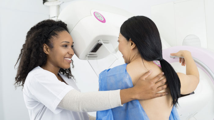
Mammography
Typically used for the early detection of breast cancers and diseases, mammograms are very low-dose x-rays specifically designed to examine the breasts. Non-invasive mammograms can detect suspicious masses and calcifications before they can be discovered by hand.
Learn more.
Learn more.
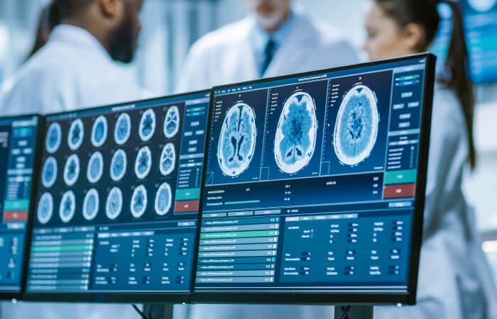
PET/CT
Positron Emission Tomography (PET) studies metabolic activity and body function, while Computed Tomography (CT) provides detailed anatomy and structure. Combined, these technologies help physicians diagnose many diseases, such as Parkinson’s Disease and Huntington’s disease, before they become apparent with other imaging examinations. With PET/CT, both exams are performed at the same time and can be used to pinpoint, for example, both the location of cancer and its growth rate. It can also be used to help diagnose the extent of a stroke or Alzheimer’s disease, and to assess heart function.
Our PET/CT’s extremely high resolution pictures give us a better chance at catching a variety of cancers in their earliest stages: whether breast, esophageal, cervical, melanoma, lymphoma, lung, colorectal, head and neck, or ovarian. Knowing more, and knowing it earlier, helps us together build the treatment plan that makes the most sense for whatever you’re facing.
Our PET/CT’s extremely high resolution pictures give us a better chance at catching a variety of cancers in their earliest stages: whether breast, esophageal, cervical, melanoma, lymphoma, lung, colorectal, head and neck, or ovarian. Knowing more, and knowing it earlier, helps us together build the treatment plan that makes the most sense for whatever you’re facing.
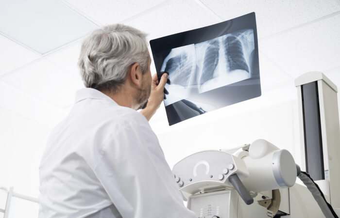
X-Rays
X-rays (or radiographs) are non-invasive and use small amounts of ionizing radiation to produce images of your internal body. The most frequently used form of medical imaging, x-rays help physicians diagnose and treat medical conditions, and are most effective for producing images of bones. The chest x-ray is the first line evaluation of many symptoms of the thorax, including cough, shortness of breath, and fever.
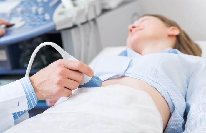
Ultrasound
Also known as sonography, ultrasound machines use high-frequency sound waves and computers to create detailed images of your internal body. Using a hand-held transducer, a physician or specialist moves the device over various parts of the body to generate very high-resolution images. Ultrasounds are especially important to monitor pregnancies and are considered safe to record the progress of both the mother and baby. Other parts of the body that are evaluated by ultrasound include the abdomen, breasts, female pelvis, scrotum, thyroid, and vascular system. In addition, breast ultrasounds are used to diagnose breast abnormalities, typically after they are discovered through a mammogram or during a physical exam. Ultrasounds are non-invasive, have no known dangers or side effects, and do not require radiation, anesthesia, or special dyes.
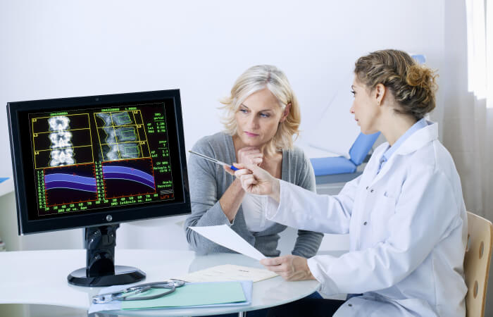
Bone Density
Bone density scanning is an enhanced form of x-ray technology. Bone density is often measured to detect or monitor osteoporosis or other medical conditions found in the bones. The scan is also known as dual-energy x-ray absorptiometry (DEXA), or bone densitometry, and is most often performed in the lower spine and hips. It is the established measuring standard for bone mineral density (BMD). In some adults and most children, the whole body is scanned. DEXA uses very low radiation amounts.
Locations

1100 E Lincolnshire Boulevard Springfield, IL 62703


400 N. 9th St. Springfield, IL 62702